For patients with hip replacements, the prospect of undergoing an MRI can raise several concerns, particularly regarding the safety and compatibility of metal implants with the strong magnetic fields used in MRI technology. While these concerns are common, the intersection of modern medical advancements and imaging technology has made MRI a viable option for most hip replacement patients.
At The Hip and Pelvis Institute, led by Dr. Nicholas H. Mast, we prioritize educating our patients on these nuanced issues, ensuring they receive the most accurate information and safe care tailored to their unique needs. This article delves into the specifics of having an MRI with a hip replacement, addressing questions seldom covered in standard discussions.
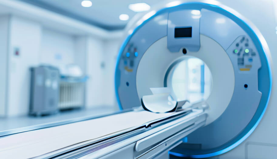
Understanding MRI and Hip Replacements
MRI, or Magnetic Resonance Imaging, is a non-invasive imaging technique that uses powerful magnets and radio waves to create detailed images of the body’s internal structures. Unlike X-rays or CT scans, MRI does not involve ionizing radiation, making it a preferred method for diagnosing a range of conditions. However, MRI’s reliance on strong magnetic fields raises questions about its compatibility with metal implants, such as those used in hip replacements.
Hip replacements typically involve the insertion of metal components made from materials like titanium or cobalt-chromium alloys. These materials are chosen for their durability and biocompatibility, ensuring they integrate well with the body while withstanding the physical demands of movement. Importantly, these metals are also non-ferromagnetic, meaning they are not attracted to magnets, which significantly reduces the risk during an MRI scan. However, the presence of metal can still cause some concerns, such as image distortion or minor heating, which must be carefully managed during the scan.
Patients with hip replacements can generally undergo MRI scans safely, but it’s crucial for the medical team to be informed about the presence of any implants. This allows radiologists to adjust the MRI settings or use specific protocols designed to minimize any potential risks, ensuring both the safety of the patient and the accuracy of the imaging results.
Is MRI Safe with a Hip Replacement?
Yes, MRI is generally safe for patients with hip replacements. The metal components used in hip implants, such as titanium and cobalt-chromium alloys, are non-ferromagnetic, meaning they do not interact with the magnetic fields used in MRI machines. This reduces the risk of complications, such as the implant moving or heating up during the scan. However, certain precautions are necessary to ensure safety.
Potential Risks and Considerations
While MRI is safe, there are potential risks and considerations to be aware of:
- Image Distortion: The metal in the hip replacement can cause artifacts or distortions in the MRI images, particularly if the area being scanned is near the implant. Radiologists use specialized techniques to minimize these distortions and obtain clearer images.
- Heating: In rare cases, the metal components may experience slight heating during the MRI. This is typically mild and does not pose a significant risk, but it is something that the MRI technicians monitor closely.
- Communication with Medical Team: It is crucial for patients to inform their healthcare providers about their hip replacement before undergoing an MRI. This allows the medical team to adjust the MRI settings and take appropriate precautions to ensure the safety and accuracy of the scan.
By taking these considerations into account, MRI scans can be performed safely and effectively on patients with hip replacements, allowing for continued monitoring and diagnosis of various conditions.
MRI Guidelines for Hip Replacement Patients
For patients with hip replacements, following specific guidelines before undergoing an MRI is essential to ensure safety and the accuracy of the imaging results. First, it’s important to inform your healthcare provider and the radiology team about your hip implant prior to scheduling the MRI. This information allows the radiologists to adjust the MRI settings and use specific protocols designed to minimize any potential risks, such as image distortion or heating.
During the MRI, patients can expect the procedure to be similar to a standard scan, but the technicians may use specialized techniques to optimize the imaging around the hip implant. It’s also crucial to remain still during the scan to avoid any movement that could affect image quality. After the MRI, the results will be carefully analyzed by your healthcare team to ensure they provide the necessary diagnostic information.
By adhering to these guidelines and maintaining open communication with your medical team, you can safely undergo an MRI with a hip replacement, ensuring both effective imaging and continued monitoring of your health.
Advances in MRI Technology and Hip Replacements
Advances in MRI technology have significantly improved the safety and efficacy of imaging for patients with hip replacements. These developments have reduced the risks associated with MRI scans and enhanced the quality of images, even in the presence of metal implants. Key advancements include:
- Metal Artifact Reduction Techniques (MARS): Specialized MRI sequences that minimize image distortion caused by metal implants, providing clearer and more accurate images.
- Improved Imaging Protocols: Tailored protocols that adjust MRI settings to account for the presence of metal, reducing the risk of heating and ensuring patient safety.
- High-Field MRI Scanners: The use of higher magnetic field strengths (such as 3T MRI) allows for better image resolution, making it easier to see around the implant and detect any issues.
These advancements not only make MRI safer for patients with hip replacements but also ensure that the diagnostic information obtained is of high quality, aiding in better clinical decision-making. As technology continues to evolve, future developments are expected to further enhance MRI compatibility with hip implants, making it an even more reliable tool for ongoing patient care.
Long-Term Monitoring and Imaging Alternatives
Regular Check-Ups and Imaging
After a hip replacement, regular follow-up appointments are crucial for monitoring the condition of the implant and the surrounding bone structure. These visits typically involve physical assessments and imaging to ensure the implant remains stable and functional. While MRI is a valuable tool for ongoing monitoring, it’s important to balance its use with other imaging modalities, depending on the specific clinical needs.
Alternative Imaging Options
For patients who may not be ideal candidates for MRI, alternative imaging techniques are available:
- X-Rays: Commonly used to assess the positioning and alignment of the hip implant, as well as to detect any loosening or wear over time.
- CT Scans: Provide detailed images of the bone and implant, useful for evaluating the integrity of the prosthesis and detecting subtle changes that might not be visible on an X-ray.
- Ultrasound: Useful for assessing soft tissues around the hip, such as muscles, tendons, and any fluid collections that may indicate complications.
By using a combination of these imaging methods, healthcare providers can effectively monitor the long-term success of hip replacements, ensuring early detection of any issues and appropriate interventions.
Conclusion
MRI scans are generally safe and effective for patients with hip replacements, thanks to advancements in technology and specialized imaging protocols. By understanding the nuances of MRI safety and following guidelines, patients can continue to benefit from this essential diagnostic tool without compromising their hip implant. Regular monitoring, combined with alternative imaging options like X-rays and CT scans, ensures that any potential issues are detected early, contributing to long-term joint health.
For personalized care and expert advice on managing your hip health, visit The Hip and Pelvis Institute or call us at (415) 530-5330 to schedule an appointment.